Leg Bone Diagram : File Human Bones Labeled - Labeled Leg Bone Diagram Clipart (#3796788) - PinClipart. In this article, we explain their function, what they are made of, and the types of cells involved. The larger bone we refer to as the tibia and is present in front of the lower leg. Bone diagram barca fontanacountryinn com. Diagram of blood and nerve supply to bone. You'll learn about the muscles, bones, and other structures of each area of the leg.
The lower leg has a structure by two bones. The femur, or thighbone, is the longest and largest bone in the human body. Human bone diagram wiring diagrams click. Visit kenhub for more skeletal system quizzes. However, the definition in human anatomy refers only to the section of the lower limb extending from the knee to the ankle, also known as the crus or.
Bones Of The Leg Photograph by Asklepios Medical Atlas from images.fineartamerica.com Time to jump right into the biggest and strongest bones in the human body. Learn how to draw the femur, patella, tibia, and fibula in this lesson! Cheek bone (zygoma) upper jaw (maxilla). However, the definition in human anatomy refers only to the section of the lower limb extending from the knee to. The foot bones shown in this diagram are the talus, navicular, cuneiform, cuboid, metatarsals. Bone diagram barca fontanacountryinn com. You'll learn about the muscles, bones, and other structures of each area of the leg. The axial skeleton and the appendicular formed by the left and right hip bones, the pelvic girdle connects the lower limb (leg) bones to the axial.
These bones are arranged into two major divisions:
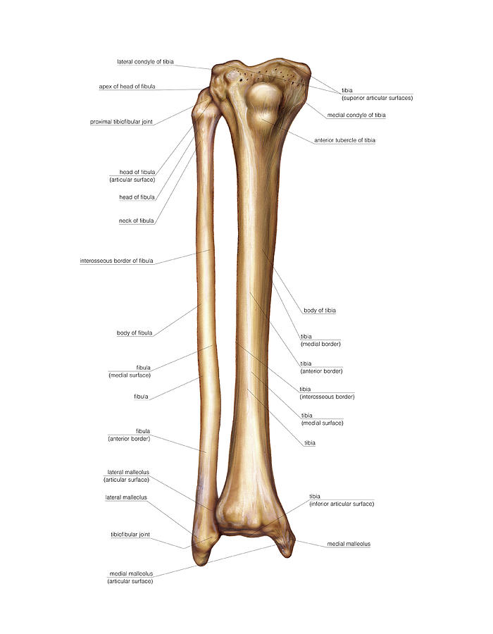
Click now to learn more about the bones, muscles, and soft tissues tibia: The bones of the leg are the femur, tibia, fibula and patella. Lower jaw (mandible) collar bone. The humerus and the femur are corresponding bones of the arms and legs, respectively. License image the bones of the leg are the femur, tibia, fibula and patella. New users enjoy 60% off. The axial skeleton and the appendicular formed by the left and right hip bones, the pelvic girdle connects the lower limb (leg) bones to the axial. The radius and ulna (bones of the forearm), shown in supination (the arm rotated outward so that the palm. Learn how to draw the femur, patella, tibia, and fibula in this lesson! The largest and most medial leg bone, forming both the knee and ankle joints. Download 2,751 bone diagram stock illustrations, vectors & clipart for free or amazingly low rates! Quizzes on human skeletal system anatomy, bone anatomy, and bone markings. Moreover, the fibula is the smaller bone that goes towards the back part of the.
Use the leg bones diagrams to learn the names of the leg bones. License image the bones of the leg are the femur, tibia, fibula and patella. Your leg bones are the longest and strongest bones in your body. In this article, we explain their function, what they are made of, and the types of cells involved. Click now to learn more about the bones, muscles, and soft tissues tibia:
Anatomy And Cell Biology 213 > Dehn > Flashcards > Thigh and leg muscles | StudyBlue from test.classconnection.s3.amazonaws.com He leg's main function in the human is for locomotion and support of the rest of the body. Time to jump right into the biggest and strongest bones in the human body. Muscles that lift the arches of the feet. The bones of the leg are the femur, tibia, fibula and patella. The human leg, in the general word sense, is the entire lower limb of the human body, including the foot, thigh and even the hip or gluteal region. Visit kenhub for more skeletal system quizzes. The foot bones shown in this diagram are the talus, navicular, cuneiform, cuboid, metatarsals. Master leg and knee anatomy using our topic page.
Health diagram bone skeleton leg knee science anchor chart human human body.
Download 2,751 bone diagram stock illustrations, vectors & clipart for free or amazingly low rates! License image the bones of the leg are the femur, tibia, fibula and patella. The bones of the leg are the femur, tibia, fibula and patella. In this article, we explain their function, what they are made of, and the types of cells involved. Cheek bone (zygoma) upper jaw (maxilla). However, the definition in human anatomy refers only to the section of the lower limb extending from the knee to. He leg's main function in the human is for locomotion and support of the rest of the body. Each leg is made up of four bones. New users enjoy 60% off. Learn how to draw the femur, patella, tibia, and fibula in this lesson! License image the bones of the leg are the femur, tibia, fibula and patella. Learn vocabulary, terms and more with flashcards, games and other study tools. Your legs are two of your most important body parts.
Master leg and knee anatomy using our topic page. They allow you to move and provide support for your upper body. You'll learn about the muscles, bones, and other structures of each area of the leg. The foot bones shown in this diagram are the talus, navicular, cuneiform, cuboid, metatarsals. These bones are arranged into two major divisions:
7.8B: Patella (The Knee) - Medicine LibreTexts from textimgs.s3.amazonaws.com Quizzes on human skeletal system anatomy, bone anatomy, and bone markings. New users enjoy 60% off. Human bone diagram wiring diagrams click. By natalia kremenon january 21, 2021in wiring diagram231 views. The human leg, in the general word sense, is the entire lower limb of the human body, including the foot, thigh and even the hip or gluteal region. Pngtree offers bone diagram png and vector images, as well as transparant background bone diagram clipart images and psd files. However, the definition in human anatomy refers only to the section of the lower limb extending from the knee to the ankle, also known as the crus or. Blood vessels and nerves enter the bone.
The foot bones shown in this diagram are the talus, navicular, cuneiform, cuboid, metatarsals.
The foot bones shown in this diagram are the talus, navicular, cuneiform, cuboid, metatarsals and calcaneus. Download the free graphic resources in the form of png, eps. The foot bones shown in this diagram are the talus, navicular, cuneiform, cuboid, metatarsals. Bones come in all shapes and sizes and have many roles. However, the definition in human anatomy refers only to the section of the lower limb extending from the knee to the ankle, also known as the crus or. These bones are arranged into two major divisions: Bones of the leg and foot, lower leg bone anatomy, leg bones anatomy, leg muscles, leg bones diagram, leg bone structure, leg anatomy muscles, parts of the lower leg. In this article, we explain their function, what they are made of, and the types of cells involved. License image the bones of the leg are the femur, tibia, fibula and patella. Click now to learn more about the bones, muscles, and soft tissues tibia: The larger bone we refer to as the tibia and is present in front of the lower leg. Quizzes on human skeletal system anatomy, bone anatomy, and bone markings. They allow you to move and provide support for your upper body.
Share :
Post a Comment
for "Leg Bone Diagram : File Human Bones Labeled - Labeled Leg Bone Diagram Clipart (#3796788) - PinClipart"


 - PinClipart)
Post a Comment for "Leg Bone Diagram : File Human Bones Labeled - Labeled Leg Bone Diagram Clipart (#3796788) - PinClipart"